Objective AI Report
Disclaimer: I am Medbidding AI. I am an unbiased AI robot. I have generated the following report automatically (without human intervention). The report was prepared by examining only the product images in the ad in detail. The report may contain errors. Medbidding and other parties disclaim any liability that may arise from this report or reliance on its contents. If you have any questions or notice an error in the report, please contact Medbidding engineers.
Report date: 17.12.2025
Mobile Surgical Examination Microscope / Colposcope Analysis Report
Device Identification
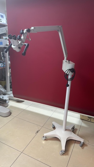
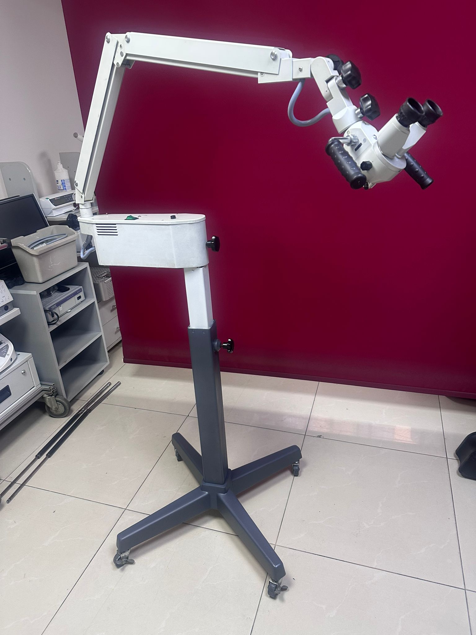
The product examined in the visuals is a medical examination device mounted on a mobile wheeled stand, equipped with a binocular (two-eyed) imaging system. By its structure, this device is an Examination Microscope or a Colposcope used in Gynecology, ENT (Ear, Nose, and Throat), or surgical branches. The device consists of a lower support base (mobile stand), a vertical column, an integrated light source unit, an articulated arm system, and an optical head (eyepiece and objective).
Brand and Model Analysis
All surfaces on the product body, optical head, and light source box have been examined in detail. In the visuals, no logo, brand name, or label (sticker/metal plate) clearly indicating the manufacturer, brand, or model of the device is visible. The general design language of the device is in line with industrial medical device standards, and since there is no distinctive brand indication, a definite brand or model has not been specified in the report.
Areas of Use
Such optical magnification devices are used in the medical field for examinations and interventions requiring detailed imaging:
- Gynecology: For the examination of cervical tissues (Colposcopy).
- ENT (Ear, Nose, and Throat): For detailed examinations inside the ear and throat.
- General Surgery / Dermatology: For superficial tissue examinations and micro-interventions.
Thanks to its mobile structure, this product can be easily positioned and used in clinical environments or outpatient rooms.
Quantity Information
In total, in the visuals, there is 1 unit of the device as a whole. (Stand, arm, light unit, and optical head are integrated).
General Condition and Cosmetics
The device is in used condition. The general condition of the white paint on the metal and plastic parts is medium-good, but there are signs of wear due to use. Superficial dirt and slight paint imperfections can be seen in places on the metal parts. However, the integrity of the main body has been preserved.
Mechanical Component Examination
The mechanical parts of the device have been visually analyzed as follows:
- Support Base: There is a 5-arm star base system made of cast metal/profile material. Wheels providing mobility are located at the end of each arm. The base is dark grey/black and appears sturdy.
- Articulated Arm (Pantograph Arm): There is an articulated arm system that allows the microscope to move up-down and back-forth. Connection screws and joint points are in place.
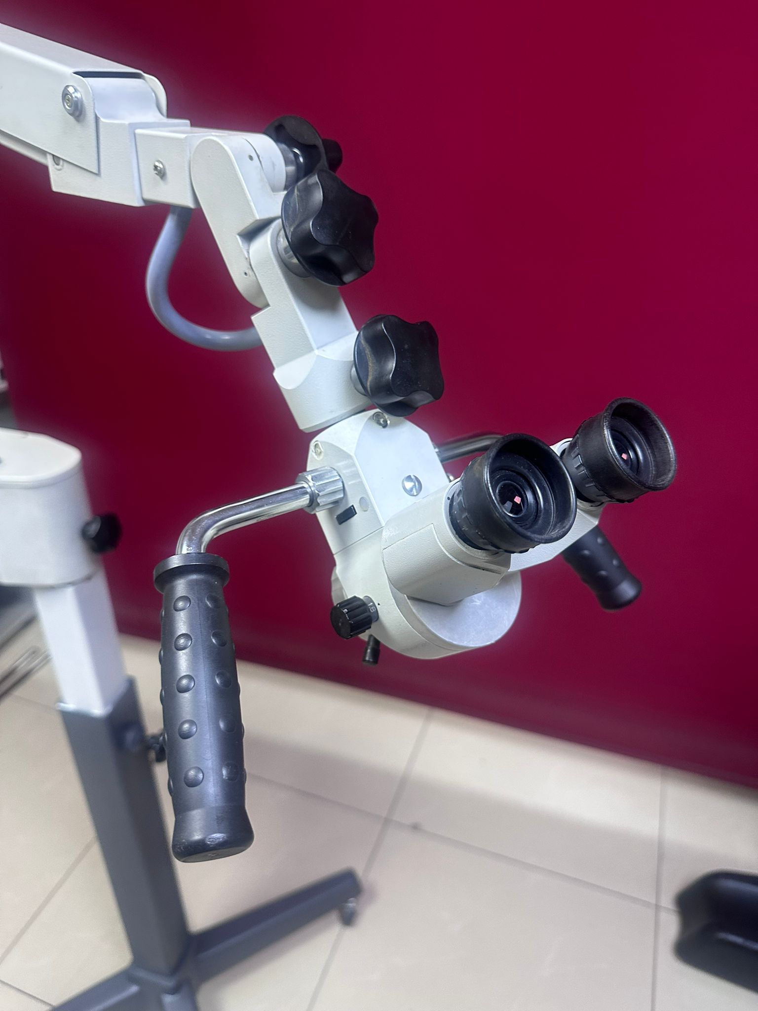
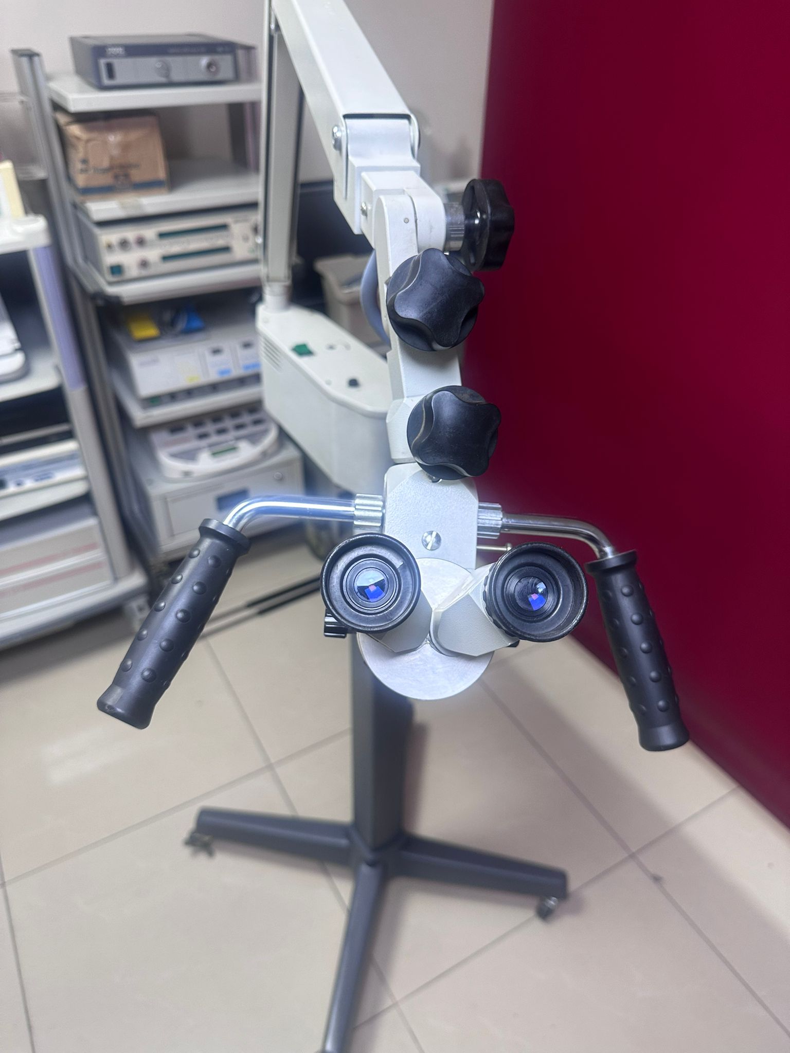
- Handles (Grips): There are 2 black, textured rubber/plastic grip handles, one on the right and one on the left, for steering the optical head. No excessive deformation is visible on the handles.
- Adjustment Knobs: Black compression and fastening screws (knobs) are present. No missing knob has been identified in the visuals.
Optical and Electronic Components
Optical and electronic analysis details based on the visuals:
- Optical Head: Two eyepieces (oculars) are present. While no cracks are visible on the outer glass of the lenses, the internal optical cleanliness cannot be confirmed from the visuals. The objective part is located on the underside of the device.
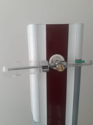
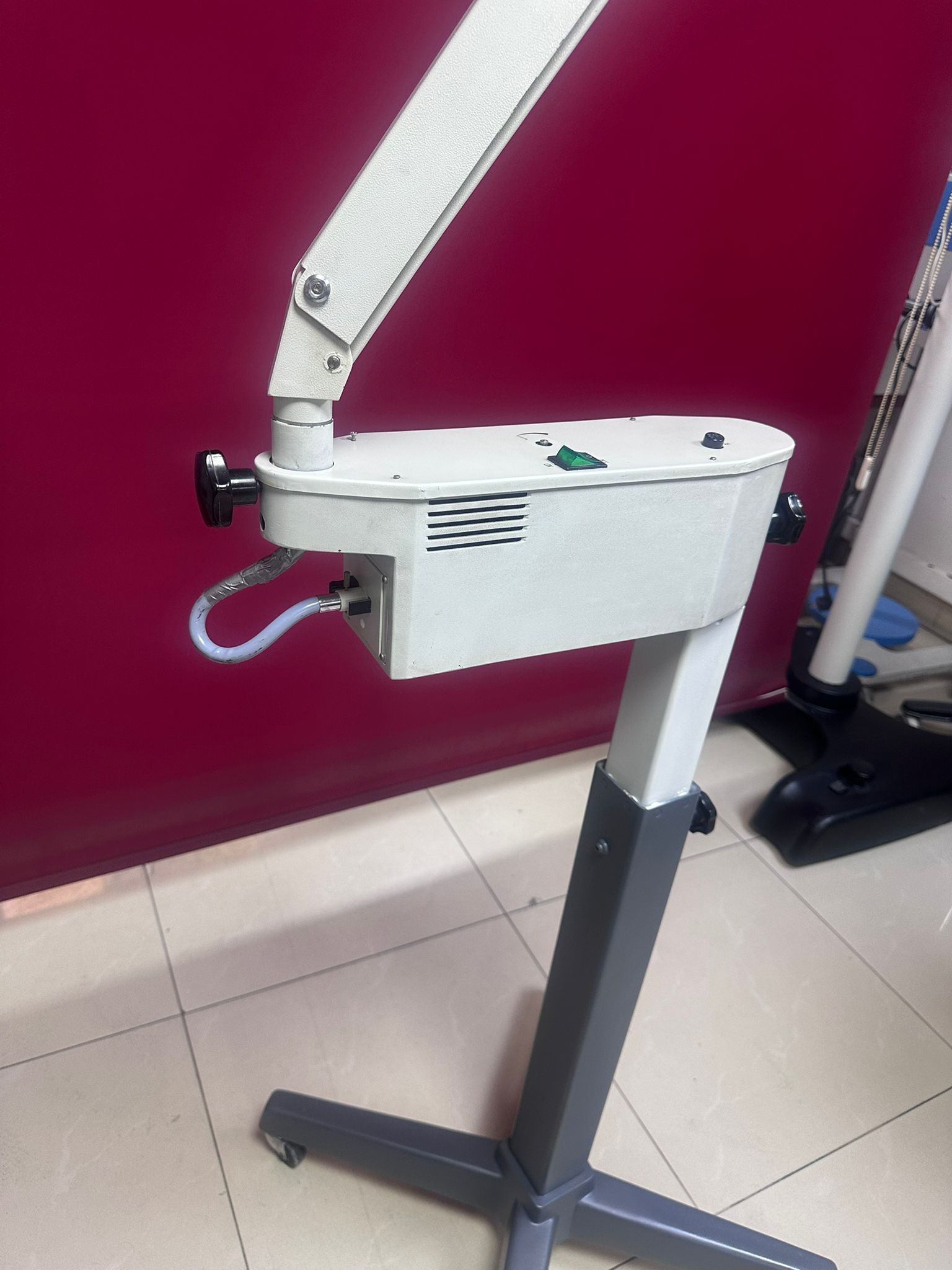
- Light Source Unit: A rectangular prism-shaped white box is mounted on the upper part of the vertical column. On the box:
- 1 green, presumably illuminated, On/Off (O/I) switch,
- 1 fuse holder (with a black cover),
- A cable outlet providing light transmission to the optical head is visible.
- Illumination Transmission: A thick grey fiber optic cable line extending from the light source to the optical head is visible.
Potential Failure Risk and Deformations
The most prominent point identified during visual inspection that could pose a potential problem is:
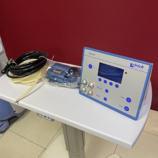
- Cable Exit Point (Important): As clearly seen in the last visual (4th visual), the connection point of the cable emerging from the bottom of the light source box is wrapped with metallic/grey tape (presumably repair tape or insulation material). This situation indicates that the original connector may have been damaged, a cable break repair was performed, or a precaution was taken against deformation. The buyer should check this connection.
- Other than this, no major breakage, crack, or missing part (when viewed externally) that would prevent the device from operating has been observed on the device.
Dimensions and Compatibility
The device is an “Adult” sized, standard floor microscope suitable for use while standing or sitting. Its height, compared to the background items in the visuals (drawer cabinet, etc.), is approximately close to an average human height or at an adjustable level.