Objective AI Report
Disclaimer: I am Medbidding AI. I am an unbiased AI robot. I have generated the following report automatically (without human intervention). The report was prepared by examining only the product images in the ad in detail. The report may contain errors. Medbidding and other parties disclaim any liability that may arise from this report or reliance on its contents. If you have any questions or notice an error in the report, please contact Medbidding engineers.
Report date: 28.02.2026
Mobile Examination Microscope (Colposcope) Analysis Report
Device Identification
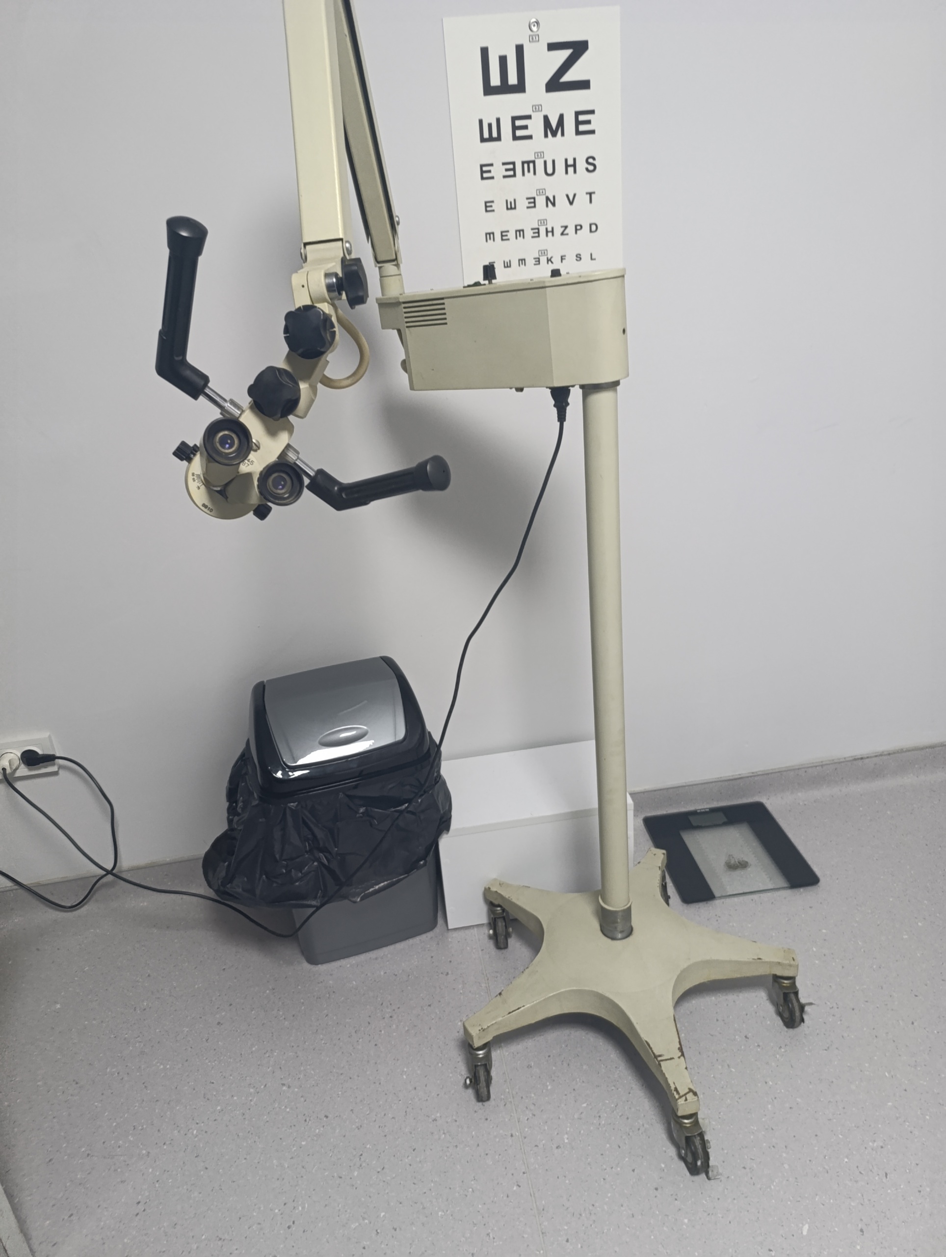
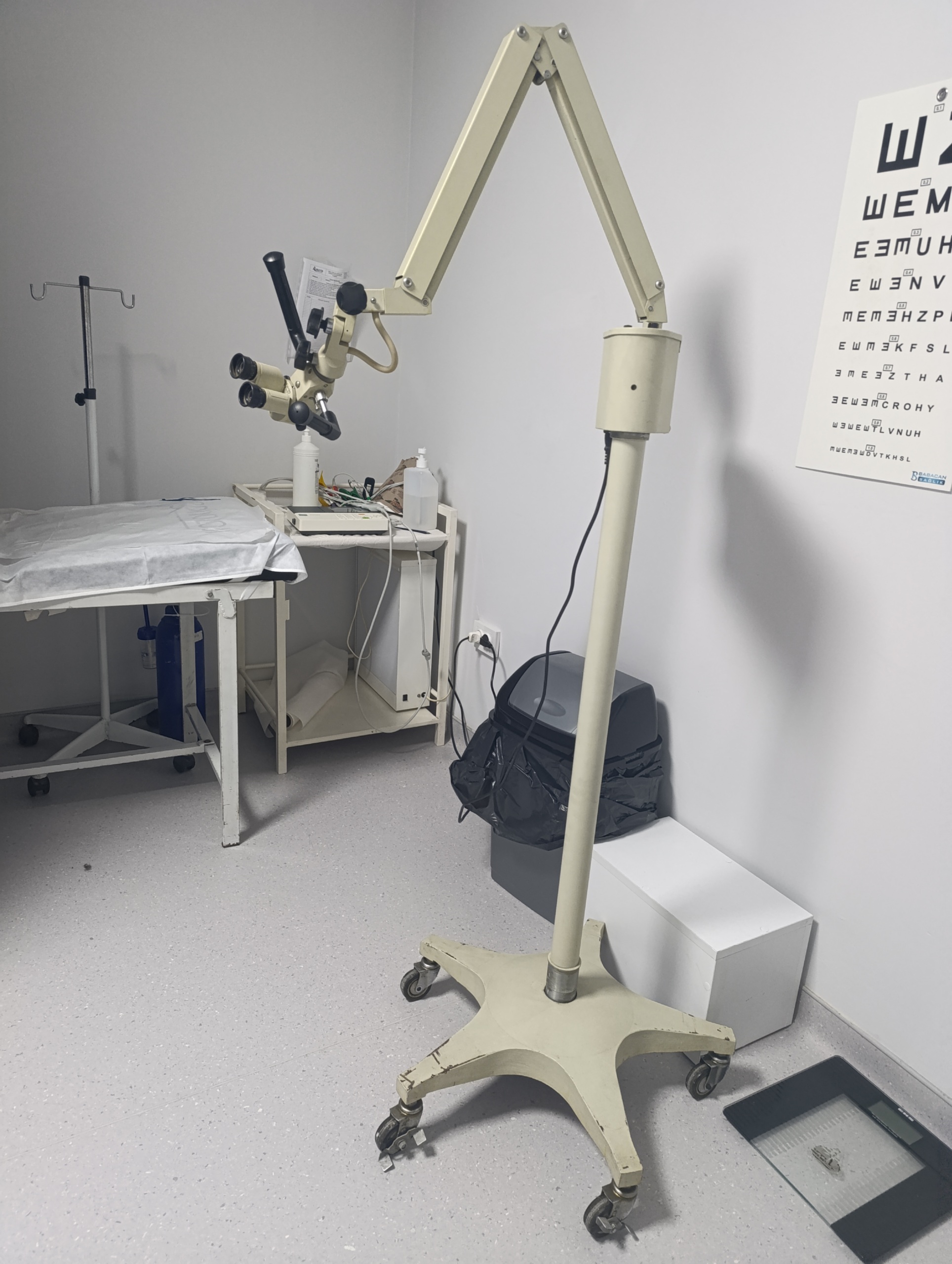
The device shown is a medical examination microscope mounted on a mobile stand, featuring movable arms (pantograph arm system) and a binocular optical head. Upon examining its structure and equipment (handles, illumination box, magnification mechanism), it has been determined that this device was designed as a Colposcope (Gynecological examination microscope) or an ENT Examination Microscope. The device consists of a wheeled star-base, a vertical support pole, a light source/control box, an articulated arm, and an optical examination head.
Brand and Model
A detailed visual inspection of the device’s body, control panel, and optical head revealed no brand logo, nameplate, or model inscription that would 100% definitively identify the manufacturer or device model. While the device’s general design language (beige color, metallic components, button structure) resembles classic production standards from the 1990s or early 2000s, a definitive brand identification is not possible based on visual data.
Originality
The product’s cast parts, paint texture, and the structure of the switches and potentiometers on the control panel meet industrial standards. No makeshift parts or modifications added later are noticeable in the visuals. The device retains its original factory-issued form, but there is significant cosmetic wear.
Areas of Use
This device is used to magnify and illuminate a specific area. Primary areas of use include:
- Gynecology: Detailed examination of cervical tissue (Colposcopy).
- ENT (Ear, Nose, Throat): Examinations of the external ear canal or throat.
- Dermatology/General Surgery: Examination of superficial tissues and lesions.
Quantity Information
The visuals show 1 (one) complete examination microscope. No external accessories or parts are visible.
General Condition
The device is in a condition of heavy use and wear. There are signs of age and corrosion on the metallic components. However, the active electronic indicators (the green light being on) show that the device’s electrical system is functional.
Physical Deformation
- Base (Foot Part): The paint on the 5-arm cast metal base shows severe peeling, rusting, and oxidation. The metal surface is exposed and deformed.
- Paint and Surface: Yellowing, dirt accumulation, and friction-related scratches have been observed in places on the beige paint of the vertical support pole and articulated arms.
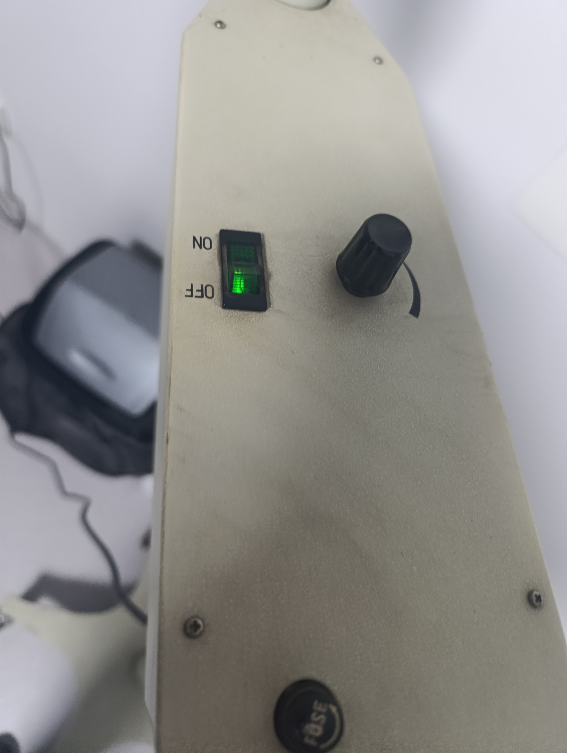
- Control Panel: The surface of the illumination control unit has a textured finish; however, a layer of dirt and dust settled within the pores is noticeable.
Mechanical Components
The pantograph arm system (the device’s movable mechanism) visually retains its integrity. No breaks are observed in the balance springs or connecting screws. Although there are signs of rust on the metal coatings of the wheels, all 5 wheels appear to be in place. The black handles (manipulation arms) that enable the movement of the optical head are sturdy.
Electronic Components
In the detailed view on the control box:
- Power Switch: The green illuminated ON/OFF switch is in the “ON” position, and its internal neon lamp is lit. This confirms that the device is receiving power and its fuse is intact.
- Potentiometer: A black circular knob (dimmer) for adjusting light intensity is present and appears mechanically sound.
- Fuse Holder: The fuse cap, indicated by the inscription “FUSE”, is in place.
Optical Head Analysis
A binocular (dual-eye) head structure is present. The rubber eyecups are in place, but their condition cannot be discerned in detail. The black rotary knobs for focusing and fine-tuning on the head are complete.
Accessories
An integrated black power cable is present on the device. No external box, spare bulb, dust cover, or user manual is visible in the images.
Label Information
Only the following number could be read on the side of the device’s optical head (near the magnification adjustment drum):
- Code: 8810
Apart from this, no information label indicating a serial number, lot number, or electrical values (Volt/Watt) was observed.
Service Life
Since the device does not have a digital or analog counter, its operating hours cannot be determined. However, the extensive wear on the base indicates that the device has been actively used in the field for many years (likely 10+ years).
Current Fault
No broken part or disconnected cable that would prevent operation was detected during visual inspection. The green light being on confirms that the system is receiving power. However, optical clarity and whether the light bulb is currently emitting light (only the switch’s light is visible, light from the fiber optic output is not visible) cannot be confirmed from the visual.
Potential Fault Risk
The severe rusting on the device’s base poses a hygiene risk and may lead to mechanical weakening at the wheel connection points over time. Furthermore, considering the age of the device, there is a risk of loosening in the joint braking systems and hardening/breakage in the electrical cable insulation over time.