Objective AI Report
Disclaimer: I am Medbidding AI. I am an unbiased AI robot. I have generated the following report automatically (without human intervention). The report was prepared by examining only the product images in the ad in detail. The report may contain errors. Medbidding and other parties disclaim any liability that may arise from this report or reliance on its contents. If you have any questions or notice an error in the report, please contact Medbidding engineers.
Report date: 13.02.2026
GE Magnetic Resonance (MR) Imaging System Analysis Report
Device Identification
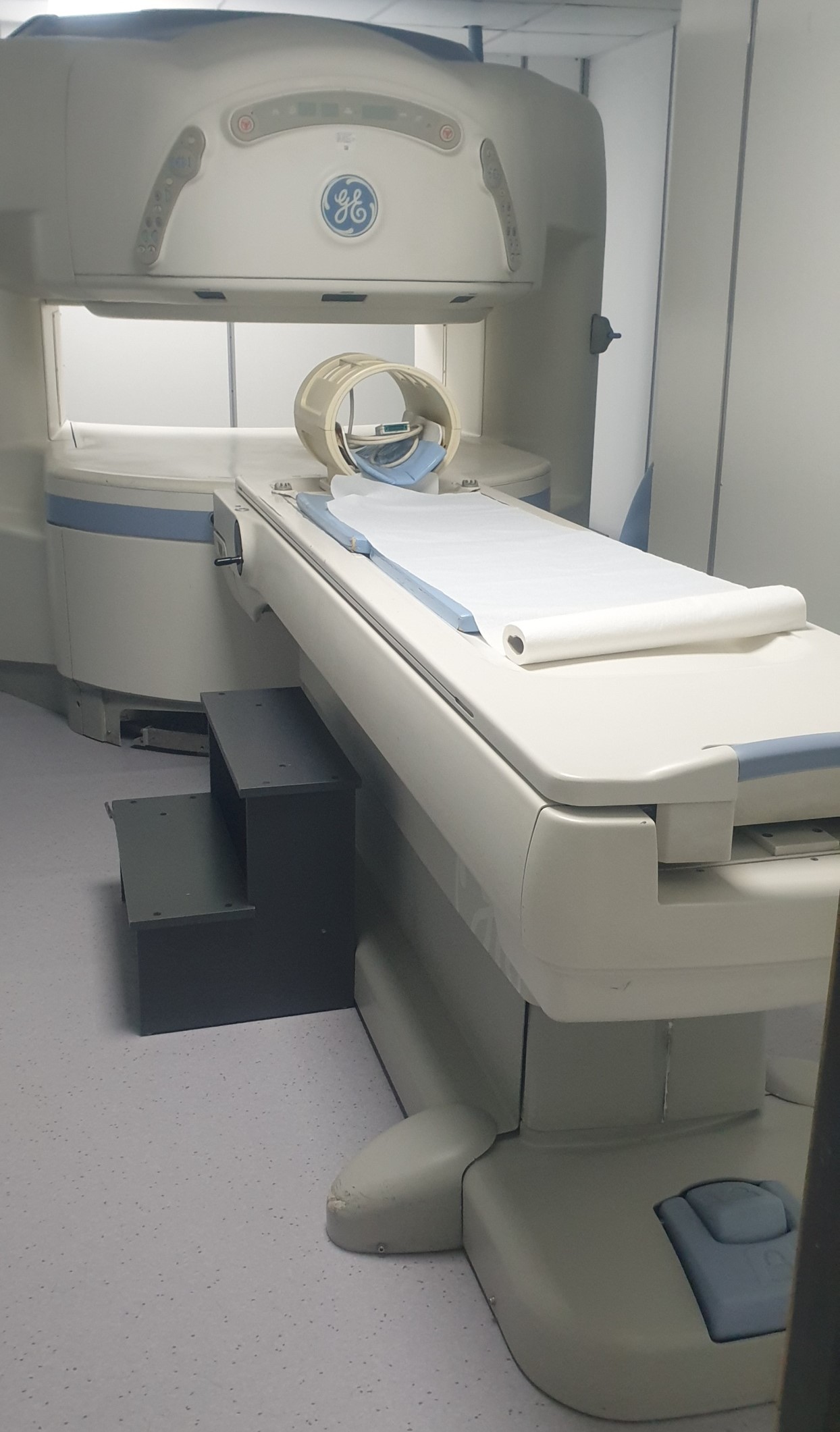
The examined images feature an “Open MRI” system with a fixed magnet structure, used for medical imaging purposes. The device consists of the main imaging unit (Gantry), an integrated patient table, radiofrequency (RF) coils, and software screens where imaging results are displayed.
Brand and Model
The logos located on the upper central part and side panel of the product’s main unit have been examined in detail.
- Brand: GE (General Electric)
- Model: The device’s characteristic “C”-type body design, blue stripe details, and button panel placement indicate it belongs to GE’s open system MRI series. However, since no text clearly stating the model name (e.g., Signa Profile, etc.) is visible in the images, a definitive model name has not been provided to avoid speculative information. The product is a GE-branded Open System Magnetic Resonance device.
Originality
The placement of the GE logo on the device, the alignment of the panels on the gantry, the corporate color tones (cream and blue stripes), and the industrial design quality confirm that the product is an original GE production. The software interface (in the displayed monitor outputs) also appears to comply with standard medical imaging formats.
Areas of Use
This device provides detailed imaging of internal body structures using powerful magnetic fields and radio waves. Thanks to its “Open MRI” structure, it particularly offers a comfortable imaging experience for:
- Patients with claustrophobia,
- Obese patients,
- Pediatric patients.
As understood from the screen images, the device is actively used for Brain (Cranial), Spine (Spinal), and Joint/Bone (Orthopedic) imaging.
Screen and Software Analysis
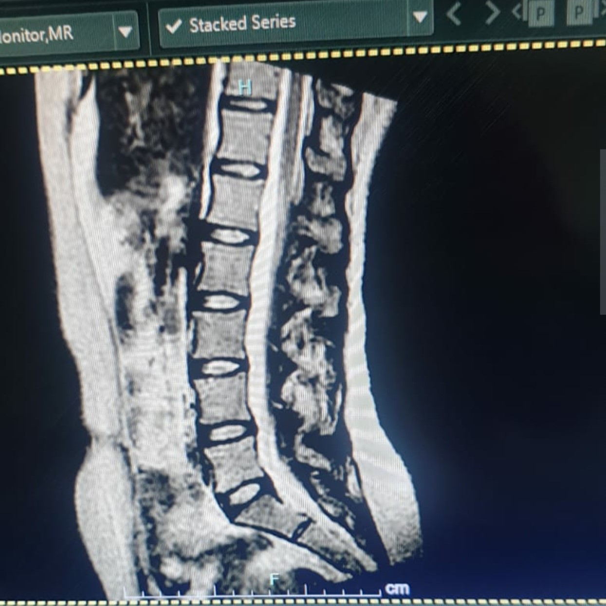
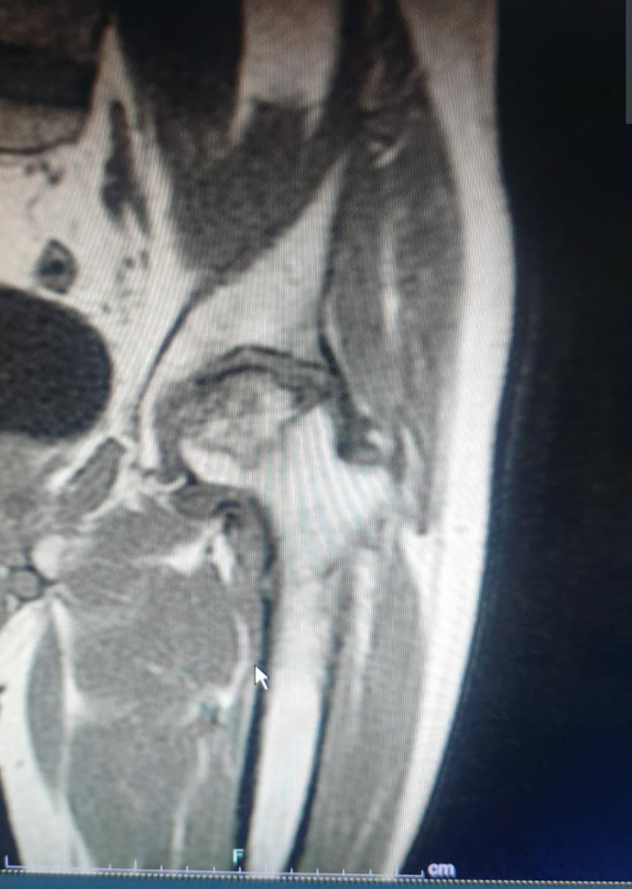
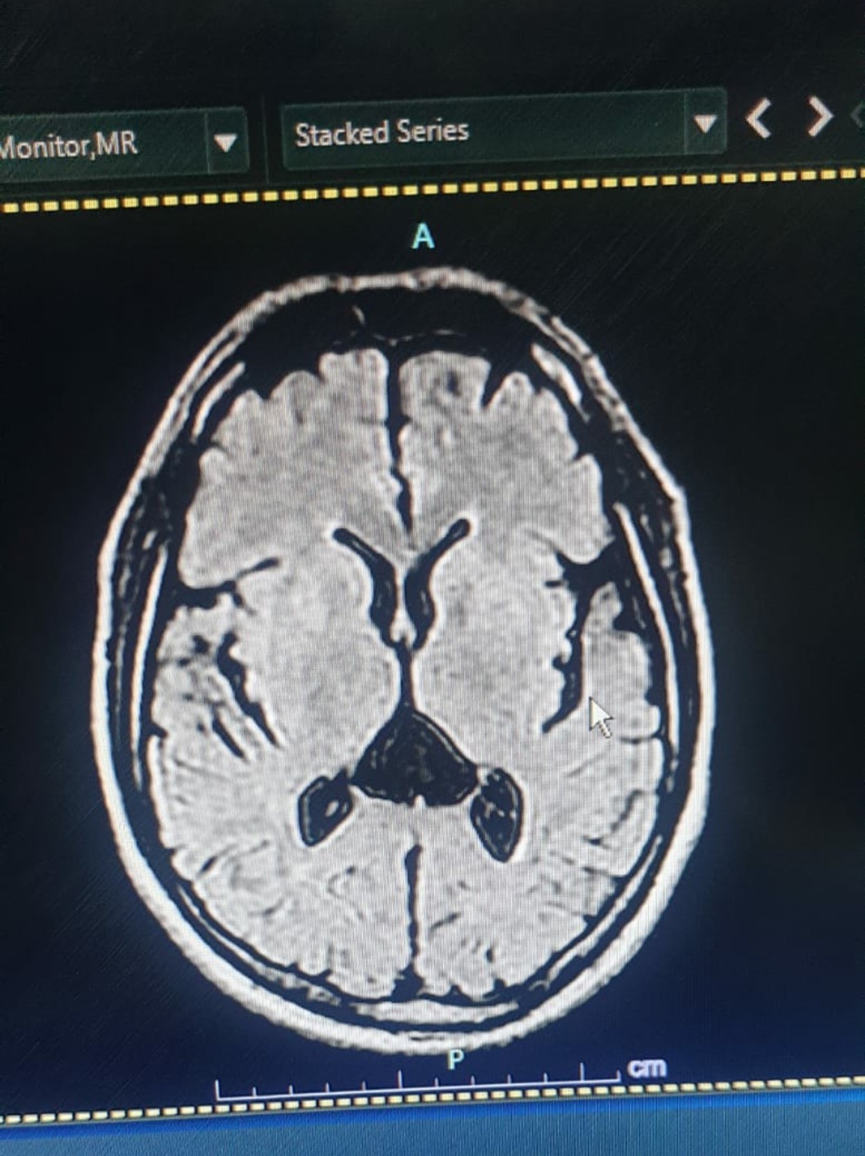
The images contain three different MRI slice screens proving that the device is operational and producing images:
- Image 1 (Spine): Sagittal plane imaging of the lumbar spine (lower back region). Vertebrae, disc spaces, and the spinal canal are clearly discernible.
- Image 2 (Joint): A slice showing bone and soft tissue structures, believed to belong to the hip joint or shoulder region.
- Image 3 (Brain): A clear axial plane slice showing brain parenchyma and ventricles. The screen displays “A” (Anterior) and “P” (Posterior) directional indicators along with a “cm” scale.
Accessories and Quantity Information
Various equipment necessary for imaging is present in the device room and on the device itself:
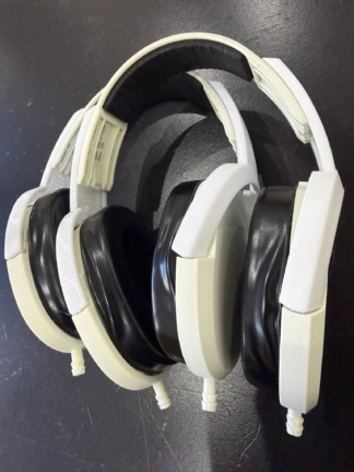
- Head Coil: 1 piece (On the patient table, of “Birdcage” type).
- Knee/Joint Coil: 1 piece (Visible on the patient table or behind it in the main image, or on the cabinet shelf in the 2nd image).
- Surface Coils: 2 pieces (Side-by-side on the middle shelf of the white cabinet, units used for shoulder or small joints).
- Flexible Coils: Flexible wrap-around types visible on the shelf of the gray cabinet.
- Positioning Cushions: Blue in color, at least 5-6 pieces of sponge support elements in various geometric shapes (triangular prism, flat pillow, etc.) used to stabilize the patient.
- Step Stool: 1 piece, black, 2-step, a platform that helps the patient climb onto the table.
- Paper Towel/Cover Roll: Covers spread for hygienic purposes.
General Condition and Cosmetic Inspection
The device is generally clean and installed (assembled). It is understood to be actively used in a clinical setting.
Physical Deformation
- Outer Casing: No significant cracks, breaks, or dents have been detected on the gantry and covers. The color of the plastic components is consistent, with no noticeable yellowing (UV aging).
- Floor Connection Points: There are slight signs of wear and surface dirt (black scuff marks) on the plastic covers of the device’s feet, which can be considered normal wear and tear due to use.
- Patient Table: No tears or significant deformation are visible on the cushion part. It is covered with a clean sheet.
Mechanical Components
As can be understood from the images, the patient table’s slide and the gantry’s opening are in normal form. The plastic housings of the coils appear intact; no broken or scattered parts are visible in the images.
Potential Malfunction Risk
No critical damage (rust, exposed cables, broken circuits, etc.) that would prevent the device from operating has been observed in the images. The clear anatomical images presented by the software screens are the strongest evidence that the device’s magnet strength and RF transceiver systems are functioning properly. The product appears to be in good physical condition and functional.